Corneal Cross-Linking for Keratoconus

Corneal Cross-Linking Procedure
Have you heard of pediatric keratoconus or corneal ectasia? Corneal ectasia refers to a group of eye diseases that cause the cornea to become thin and bulge. Keratoconus is one of those eye diseases, and it can result in blurry vision, glare, halos around lights, sensitivity to bright light and eye irritation. It often begins in the teenage years, especially if there’s a family history of the condition, but some people don’t notice symptoms until their 20s or 30s. Our cornea center at Marietta Eye Clinic offers Corneal Cross-Linking eye surgery to help stop the progression of keratoconus.
What Is Keratoconus?
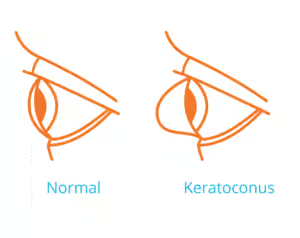
Keratoconus is an eye disease that affects the shape of the cornea. It’s due to a problem with the collagen in the eye, which is a specific type of protein. Collagen is responsible for providing structure and strength to various components in your body. You can think of it like the glue that holds certain cells together so that they can function. This protein can be found in your muscles, bones, skin and connective tissues, and it’s estimated that 30 percent of the human body’s proteins are collagen.
The collagen in the eye is known as type V. Type V is also found in the skin and hair. When the collagen starts to break down, it results in a loss of structure. For individuals with keratoconus, it means that the cornea becomes thin and starts to bulge outward. This can give the cornea a cone-like appearance, which is how this condition got its name.
When Does Keratoconus Occur?
It’s estimated that keratoconus occurs in one person out of every 2,000 . Noticeable symptoms typically start between the ages of 13 and 30. As a parent, you may be able to spot some signs of the condition. For example, if your child was once good at sports and suddenly struggles to catch the ball or has difficulty reading the whiteboard at school, it might be due to keratoconus. Of course, if you or your children notice any changes in vision, it’s time to see an eye doctor.
Keratoconus Symptoms and Causes
When the cornea becomes thin and starts to bulge, light doesn’t enter the eye correctly, so it isn’t focused properly on the back of the eye. The exact causes of why the collagen in the cornea breaks down is unknown. Symptoms of this eye disease usually become noticeable in high school. The first symptom is usually blurry vision, and it’s very important that the individual have their eyes tested as soon as they notice a change in their vision. Comprehensive eye exams can detect changes in the cornea as well as other problems with the eyes.
Additional symptoms of Keratoconus include:
- An increase in glare and seeing halos around lights, especially at night
- A sudden increase in sensitivity to bright lights
- Vision appearing cloudy
- Worse vision over time
- Eye irritation and/or pain
Keratoconus Risk Factors
While the cause of keratoconus is unknown and there’s no way to prevent it, there are some risk factors that could indicate an increased risk of developing the condition.
- Family history of keratoconus
- Experiencing chronic eye inflammation
- Constantly rubbing the eyes
- Having Down Syndrome, as it is estimated that between 5 and 30 percent of individuals with Down Syndrome develop keratoconus
Stages Of Keratoconus
There are four stages of keratoconus. In order to treat the eye disease and achieve the best possible outcome, it’s important to get it treated in the early and moderate stages with Corneal Cross-Linking surgery, which can help stop the progression and prevent the need for a corneal transplant.
Early
The early stage of keratoconus is characterized by slight distortions in the cornea. Most people don’t notice any changes in their vision, and if there are changes in the vision, they can be corrected via glasses and toric contact lenses.
Moderate
The moderate stage is sometimes grouped with the early stage. This is because the changes in the cornea progress, but the individual may only notice slight changes in their vision, like the words on the whiteboard at school looking blurry. The good news is that moderate pediatric keratoconus can still be corrected via contact lenses. However, the recommended lens type changes from soft/toric to hybrid, which contains a hard center and soft edges.
Advanced
Corneal scarring starts to occur in the advanced stages. This can cause the cornea to become cloudy, leading to increasingly blurry vision, sensitivity to light and eye pain. Scleral lenses can help correct vision at this stage. However, this is also the stage that starts the conversations about corneal transplants.
Severe
The advanced and severe stages are often grouped together due to the progressive corneal thinning and scarring. As the eye disease progresses, contact lenses may become too uncomfortable to wear and not provide the desired amount of vision correction. At this stage, corneal transplants are often recommended.
Corneal Cross-Linking
Corneal Cross-Linking (CCL) eye surgery can help slow or stop the breakdown of the collagen in the cornea. At Marietta Eye Clinic, our ophthalmologist, Mark Morel, M.D., performs the iLink CCL surgery.
iLink CCL
iLink is the only FDA-approved procedure to help slow or stop the progression of keratoconus. It is considered an epi-off surgery. This treatment utilizes UV light from the KXL system and Photrexa® ️ Viscous eye drops to slow or stop the progression of pediatric keratoconus by strengthening the links in the collagen, called collagen fibrils. CCL is an outpatient procedure and takes about an hour.
What To Expect During Your Corneal Cross-Linking (CCL) Eye Surgery
The procedure starts with the administration of medication to help you feel relaxed as well as numbing eye drops to help ensure that you do not feel any pain. It’s important to note that you will be awake for the entire iLink CCL procedure, which typically takes about an hour. However, you should expect to spend at least two hours at the clinic.
After the numbing eye drops and relaxing medication have taken effect, our eye surgeon, Dr. Mark Morel, will remove the epithelium, which is the thin layer that covers the cornea. This is what makes this procedure an epi-off, and the layer is removed in order to ensure that the riboflavin eye drops successfully penetrate the tissue of the cornea.
Riboflavin Eye Drops
Once that epithelium has been removed, our eye surgeon will apply the Photrexa® ️ Viscous eye drops, which are comprised of riboflavin 5’-phosphate in a 20 percent dextran ophthalmic solution. The Photrexa® Viscous eye drops are left in your eyes for about 30 minutes so that they can penetrate the cornea.
Ultraviolet (UV) Light
After 30 minutes have elapsed, our eye surgeon then shines a UV light into your eyes. This step also takes about 30 minutes, and during the UV light procedure, additional drops are put into your eyes.
Combining Riboflavin and UV Light
The combination of the riboflavin drops, and UV light helps increase the cross-linking of the collagen in your cornea, which helps slow or stop the progression of keratoconus. It’s important to note that it does not restore vision that’s already been lost. Once the procedure is complete, a contact lens will be placed over the treated eye in order to prevent debris from entering the surgical site and to aid in healing.
What to Expect After CCL Surgery
Once the CCL surgery is complete, you may feel some eye discomfort and sensitivity to light. This is perfectly normal. Our eye surgeon will give you a sheet of post-operative instructions to help you understand what to expect and what to watch for. We do recommend that you rest for the remainder of the day and avoid rubbing your eyes. Rubbing your eyes could dislodge the contact lens.
Our eye doctor may also recommend an OTC medication to help alleviate any eye discomfort and prescribe antibiotic eye drops to help prevent infection. You may also want to avoid bright lights and wear sunglasses when outside. Most individuals can resume the majority of their daily activities the next day. However, any activity, like sports, should not be participated in until the eyes have fully healed, which can take several weeks. Once your eyes have fully healed, which can take up to two months, our eye doctor will fit you with contact lenses or glasses.
How effective is the procedure?
Corneal Cross-Linking (CCL) eye surgery is 95 percent effective in hauling the progression of keratoconus. According to our eye surgeon, Dr. Mark Morel, “corneal cross-linking strengthens the corneal tissue, stabilizing the cornea and preserving the vision for patients with progressive keratoconus. It’s a proactive intervention tailored especially for those who are experiencing an ongoing worsening of their keratoconus or corneal ectasia.”
What are the long-term outcomes?
According to a study on the Five ‐ year corneal cross ‐ linking outcomes: A Save Sight Keratoconus Registry Study , the procedure is effective at stabilizing the cornea for at least five years. However, the American Academy of Ophthalmology reports that CCL procedures result in benefits for up to 10 years. The good news is that CCL can be repeated in the future as long as the corrected distance visual acuity (CDVA)is still within acceptable parameters.
Insurance Coverage for Corneal Cross-Linking?
Ilink Corneal Cross-Linking is covered by the majority of insurance plans. Mark Morel states,
“Here in Georgia, due to the medical necessity of Corneal Cross-Linking, commercial insurance coverage is now more than 95 percent.”
It’s important to note that insurance typically only covers Corneal Cross-Linking procedures that are FDA-approved and use the Avedro medications and devices, of which iLink is a part of. It does not typically cover epi-on procedures or procedures that are not FDA-approved. If your medical insurance provider is one of the 5 percent that doesn’t cover CCL, other options may be available, including appealing to your current insurance provider and opting for financing.
Is Cross-Linking Right for Me?
Corneal Cross-Linking is typically recommended for individuals with unstable keratoconus or progressive keratoconus. It is not recommended for patients whose corneas are stable. Therefore, it’s very important to monitor the progression of the corneal changes. If the individual notices progressively blurry vision, trouble seeing at night and eye pain, CCL may be right for you. After all, early intervention helps improve long-term outcomes.
If you’ve noticed a deterioration in your vision or your child has said that they can’t see as well, it’s time for a consultation with our CCL ophthalmologist, Mark Morel. Schedule an appointment or call us at 770-427-8111.
Cornea Crosslinking Doctors
 Dr. Mark Morel is our corneal specialist for the CCL surgery at Marietta Eye Clinic. He graduated from the Emory University School of Medicine and completed a residency and fellowship at Emory University. He performs ongoing research into Neurotrophic Keratitis and Ocular Cicatricial Pemphigoid, and he wants you to know that
“If you or a loved one has progressive keratoconus, you don’t have to face it alone. We urge you to take the first step towards safeguarding your vision by scheduling a consultation with us today.”
This is because,
“Without intervention, it may lead to significant visual impairment.”
The good news?
“Corneal Cross-Linking is a minimally invasive procedure that we offer right here at Marietta Eye Clinic that can halt the progression of keratoconus.”
Contact Us
Ready to have a conversation about improving your eyesight? At Marietta Eye Clinic, we’re dedicated to delivering comprehensive eye care combined with exceptional customer service. Let’s get started.