The Routine Eye Exam and What it Entails by Leonard Thurschwell, O.D.
April 29, 2020
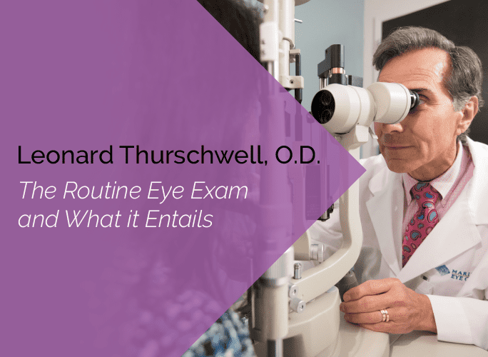
For many, vision is the sense that would be the toughest to lose. Yet despite how much people rely on their vision, many don’t get examinations as often as recommended. Regular exams allow eye doctors to detect abnormalities before they become irreversible problems. For people in high risk groups, such as those living with systemic health problems that affect the eyes, regular exams are even more important to maintaining ocular health.
When done properly, eye exams require that the patient be in the doctor’s office between 60 and 90 minutes. Many patients question why eye exams take so long, especially if they’re just there to renew a prescription. A comprehensive eye exam looks at more than just refraction, though refraction is admittedly a big part of the exam.
What is Covered and Discussed During an Eye Exam?
History
- Chief complaint – In the patient’s own words, why are they presenting for an eye examination? Is this an annual eye examination? Is their vision blurred at distance, up close, or both? Do they feel they need their prescription changed, do they just want a new pair, or have they lost their most recent prescription? Are they having any problems with their eyes, such as a floater(s), double vision, trauma, or irritation? Do they wear contact lenses or have any interest in trying them? Do they have any systemic conditions such as diabetes or arthritis for which another doctor wants their eyes checked?
- Personal ocular history – Is the patient new to the practice, or are they an established patient? Previous eye exam results are reviewed for abnormalities or anomalies that were discovered in the past or conditions that are being followed such as glaucoma or a history of retinal tear or detachment. Has the patient had any ocular conditions that required treatment? Does the patient suffer from headaches or double vision? If the patient is established, then the above items are usually already documented, except for those items that occurred between their last exam and the present visit. Additionally, an eye doctor will want to know if the patient using any ocular medications, whether over the counter or prescribed.
- Personal systemic and general history – Does the patient have any health problems, such as high blood pressure, diabetes, thyroid abnormalities, or autoimmune conditions? Is the patient taking any over-the-counter or prescribed medications? Is the patient allergic to any medications, or have they had previous adverse side effects to previously used eye medications for treatment or examination? If the patient is a child, were they born premature? Have they been hospitalized for any unusual health conditions? Have they met their age appropriate growth landmarks per their pediatrician? How are they performing in school?
- Family ocular history – Do any blood relatives have ocular conditions that be hereditary? These can include strabismus (turned eye), glaucoma, macular degeneration, certain corneal conditions, and retinal tears or detachments.
- Family systemic history – Does the patient have any family members with a history of possible hereditary conditions, such as diabetes?
Visual Acuity
A visual acuity test provides the fraction that quantifies the patient’s ability to identify smaller and smaller letters, such as in the phrase “20/20 vision.” The first number, or numerator, refers to the distance from which the test is conducted — usually 20 feet — and second number, or denominator, refers to the smallest letter which is read. The smaller the denominator, the clearer a person’s vision. Thus, a person with 20/25 vision sees more clearly than one who sees 20/60. This test is usually conducted while the patient wears glasses or contacts. Visual acuity is usually checked at close (reading) levels as well.
External Testing
- Color vision is usually tested using images of colored dots that make up numbers that are a different color than the dots around them. Color vision abnormalities tend to be hereditary. They are fairly common in males (about 10%) and very uncommon in females (lower than 1%). Congenital deficiencies are usually symmetrical between the eyes. Color vision abnormalities can also develop as a result of eye disease. These color vision abnormalities have an equal probability between males and females and can be asymmetric between the eyes.
- Stereovision is usually tested by having a patient look at 3-D images while wearing polaroid glasses. Stereovision allows a person to comprehend spatial perspective, length, width, and depth. A person who has very reduced vision in one eye will usually have reduced stereovision.
- Eye alignment tells a clinician whether eyes are working together. In some instances, one eye may be viewing slightly above, below or to the side of the other eye. Small deviations are tolerated by the visual system. If muscle paralysis or weakness causes a larger deviation, double vision may be present. This level of eye misalignment may require treatment with special glasses or surgery.
Refraction
When refraction is tested, an instrument called a phoropter is placed in front of the patient’s eyes and various lens combinations are demonstrated until the clearest vision is determined. Patients will either require no vision correction or be categorized as myopic, hyperopic, astigmatic, or presbyopic. If the patient’s chief complaint includes blurred vision, a prescription will be dispensed.
The following are the most common conditions that would require someone to wear glasses or contacts:
- Myopia, also referred to a nearsightedness or shortsightedness, is the condition of having blurred distance vision.
- Hyperopia, also called farsightedness, refers to blurred vision at distance, near, or both, depending on a patient’s age.
- Astigmatism is an asymmetric curvature of the cornea or crystalline lens that can cause blurred vision at both distance and near. This is often described as a “football-shaped” eye and usually found in conjunction with nearsightedness or farsightedness.
- Presbyopia is characterized by blurred vision up close and is secondary to aging.
For those who require corrective glasses or contact lenses, there are 3 types of lenses available.
- Single vision glasses or contacts correct for vision at a single distance.
- Bifocal glasses or contacts, also referred to as conventional or progressive, correct for two distances.
- Trifocal glasses or contacts, also referred to as conventional or progressive, correct for three distances.
Slit Lamp (Biomicroscope) Exam
A slit lamp allows a practitioner to thoroughly examine the outer or external aspects of the eye, such as the lids, conjunctiva, corneal tissue, iris, and sclera (the white of the eye). If the eye is dilated, the internal structures can also be examined in great detail. Abnormalities of these structures may indicate cataracts, macular degeneration, optic nerve disease, or retinal disease.
Intraocular Pressure (Glaucoma Test)
Intraocular pressure is usually tested with an instrument attached to a slit lamp. The instrument will gently touch the patient’s anesthetized eye and determine its intraocular pressure (IOP). Normal IOPs range between 10 and 21 millimeters (mm) of mercury. Higher IOP may indicate glaucoma. Additional testing may be performed to rule out this potentially sight-threatening condition.
Ophthalmoscopy
An ophthalmoscopy is commonly performed with a handheld instrument or direct ophthalmoscope (DO) whereby the clinician approaches each eye and looks through the pupil to examine the eye’s internal structures. Whether the eye is dilated or not, this handheld instrument offers a limited, two-dimensional view of the retina. Another type of ophthalmoscope, referred to as an indirect ophthalmoscope (IO), can be worn on the clinician’s head. When the pupils are dilated, an IO allows a clinician to thoroughly examine the internal contents of the eye in 3 dimensions. Many abnormalities can be observed with an IO.
Patients often have various excuses for not wanting to be dilated — school, work, light sensitivity after dilation, blurred vision, etc. Be sure to schedule your exam when dilation can be performed. If, for some reason, dilation is not an option, there are cameras that permit a wide angle internal view of the retina that can be used instead.
Discussion of the Results
After testing is complete, the results are reviewed with the patient and a final disposition is formulated. Will a new prescription be dispensed? Is there some aspect of the exam that will require more frequent evaluations, more specific testing, or referral to a specialist within the practice? Were there any findings that require further evaluation by a general medical practitioner or specialist outside the practice?
When Should Eye Exams be Conducted?
Most children should have an eye exam by age 5, although exams can be performed sooner if needed. How a child interacts with others and learns depends on clear, unobstructed vision. Impaired eyesight can have long-term consequences for mental growth and development. Children born premature are at higher risk of visual complications. It is never too early to have a thorough visual assessment to check for problems that may cause school-related or personal problems.
Most adolescents should have an eye examination at least every 2 years if they don’t already wear glasses or contact lenses. As children grow, their eyes change. An eye doctor can determine whether more frequent evaluations are necessary to keep your child seeing well. Dilated exams can reassure the parents that the internal structures of the eyes remain healthy.
Adults should have eye exams at least every 2 years, depending on results of previous exams or whether they require vision correction. Whether or not changes in the patient’s glasses or contact lens prescription are necessary, the health-related aspects of an eye exam remain paramount. Periodic exams help determine whether a patient’s visual system remains normal. Dilation becomes more critical as people age to assess overall external and internal eye health.
Patients with systemic diseases such as hypertension, diabetes, or autoimmune diseases may require more frequent evaluations. How often these examinations are recommended depends on the severity of the systemic disease, how severe the vision loss may be at present, or the potential for initial or further vision loss.
Prioritize Your Ocular Health
Eye examinations are critical to overall health. They should be conducted periodically and perhaps more frequently if a patient presents a condition that requires close monitoring. Many people conflate a vision (glasses or contact lens) exam with a comprehensive eye exam. Glasses or contact lens prescriptions do not reflect the health of the eyes and must be accompanied by a thorough internal examination of each eye while dilated to assess ocular health. Don’t cheat yourself or your family of having the most comprehensive exam performed by a capable clinician.
More About Ocular Disease Specialist Leonard Thurschwell, O.D.
Dr. Thurschwell is a primary care optometrist with the Marietta Eye Clinic who also provides ocular surgery co-management. He earned his doctor of optometry from the Pennsylvania College of Optometry. He is a member of the American Academy of Optometry, the Georgia Optometric Association, and the American Optometric Association. He has had his research published in the Southern Journal of Optometry and the Journal of the American Optometric Association. Read more about him in his bio.
References and Additional Resources
The American Academy of Ophthalmology (AAO) is the world’s largest association of eye physicians and surgeons. It describes itself as a “global community of 32,000 medical doctors who protect sight and empower lives by setting the standards for ophthalmic education and advocating for our patients and the public.” Its website is a great resource to learn more about how to care for your eyes.
The American Optometric Association (AOA) describes itself as the leading authority on quality care and an advocate for our nation’s health, representing more than 44,000 doctors of optometry, optometric professionals, and optometry students.
Set Up A Consultation
When it comes to your eyes, nothing beats a face-to-face consultation with one of our eye doctors. Request an appointment to meet with one of our specialists.